
ECG
ECG
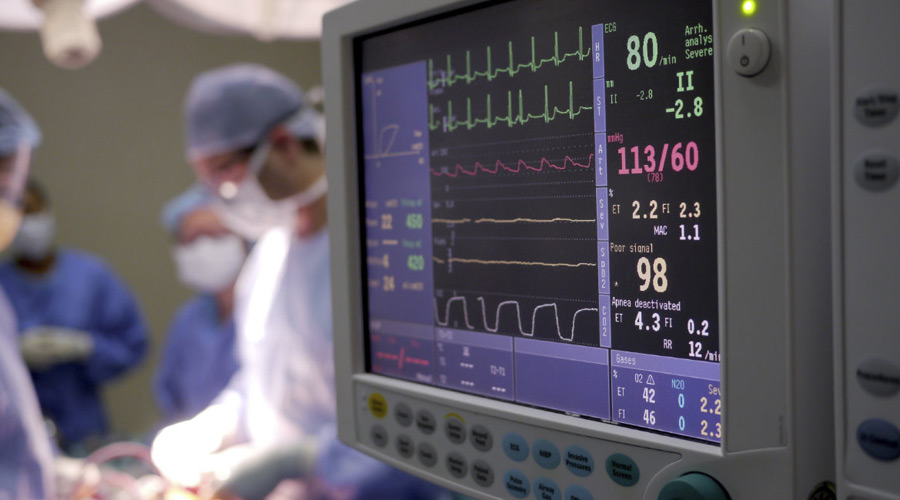
An Electrocardiogram (ECG or EKG) is a diagnostic test that records the electrical activity of the heart over a period of time. It is a valuable tool in cardiology and is used to assess the heart's rhythm and electrical conduction, identify cardiac abnormalities, and diagnose various heart conditions. Here's how an ECG works and what it can tell us:
How ECG Works:
-
Electrodes: During an ECG, small, adhesive electrodes are placed on specific areas of the skin, usually on the chest, arms, and legs. These electrodes are connected to leads that transmit electrical signals from the heart to an ECG machine.
-
Recording: The ECG machine records the electrical impulses generated by the heart as it contracts and relaxes. These impulses are transmitted as waves on the ECG tracing.
-
Tracing: The ECG tracing is a visual representation of the heart's electrical activity, displayed as a series of waveforms on a strip of graph paper or on a digital screen.
What an ECG Can Reveal:
-
Heart Rate: An ECG provides information about the heart rate (number of beats per minute) by measuring the distance between the R waves on the ECG tracing.
-
Rhythm: The ECG helps assess the regularity of the heart's rhythm. Normal rhythms are typically referred to as sinus rhythms. Irregular rhythms can indicate conditions like atrial fibrillation.
-
Cardiac Abnormalities: ECGs can detect various cardiac abnormalities, including:
- Arrhythmias: Abnormal heart rhythms, such as atrial fibrillation, ventricular tachycardia, or bradycardia.
- Myocardial Infarction (Heart Attack): ECG can reveal changes in the ST segment, T wave, and Q wave, which can indicate damage to the heart muscle.
- Cardiac Ischemia: Reduced blood flow to the heart muscle can be seen as changes in the ST segment on the ECG, often indicating angina.
-
Hypertrophy: ECG can also detect signs of left ventricular hypertrophy (enlargement of the heart's left ventricle), which can be associated with conditions like high blood pressure.
-
Electrolyte Imbalances: Disturbances in blood electrolyte levels (e.g., potassium, calcium) can manifest as ECG changes, potentially affecting heart function.
-
Medication Effects: ECG can show the effects of certain medications or toxins on the heart's electrical activity.
Types of ECGs:
-
Resting ECG: This is the most common type of ECG, done while the patient is at rest. It provides a baseline assessment of heart function.
-
Stress ECG (Exercise ECG or Treadmill Test): This test involves an ECG while the patient exercises, typically on a treadmill or stationary bike. It can help diagnose coronary artery disease and assess exercise tolerance.
-
Holter Monitor: This is a portable ECG device that a patient wears for 24-48 hours or longer to record heart activity continuously. It's useful for diagnosing intermittent arrhythmias.
-
Event Monitor: Similar to a Holter monitor, but worn for a more extended period, an event monitor is used to capture intermittent symptoms like palpitations.
ECGs are invaluable tools in diagnosing and managing heart conditions. The results are interpreted by healthcare professionals, often cardiologists, to guide treatment decisions and monitor cardiac health.